Monday, May 31, 2010

Cephalopod eyes

I just wrote a big post about cephalopod eyes, and then realized that I had neglect to show any real-life pictures of cephalopod eyes.  This blog seems to be getting a bit dry, so let's take some time off and just gaze at some of our beautiful, squishy friends.  All images are from the wikimedia commons and have been under a creative commons license.

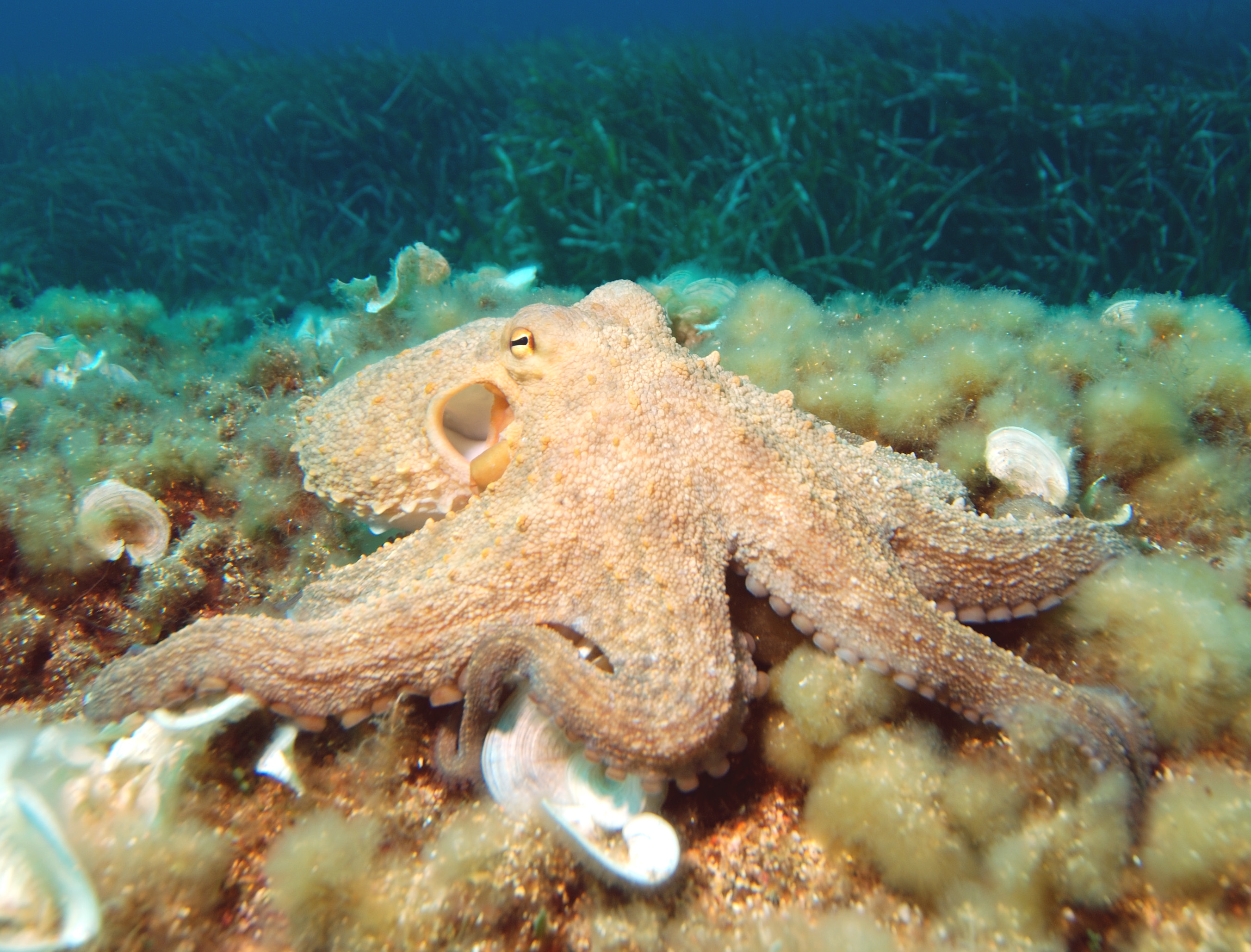
(Photo by Parent Géry)  This guy is Octopus vulgaris, also known as the common octopus.  It the most-studied octopod.  You can see the slit-shaped pupil clearly.


(Photo by Theasereje)  Here's a body shot of another O. vulgaris.  Notice how they can look at you with both eyes at the same time - they have the capability for binocular vision.  Octopuses, however, prefer monocular vision, and will always use just one eye to sight their prey during an attack (for more info, see this "Lateral asymmetry of eye use in Octopus vulgaris" by Byrne et al.)


(Photo by Elapied)  Here we have another gorgeous shot of O. Vulgaris peering out of a hideout with one eye.


(Photo from www.opencage.info)  This is an ocellated octopus, O. ocellatus.  Besides being very cute, as he peeks out from the shell, he is probably using his mostly monocular vision to monitor his whole environment for danger.


(Photo by Jens Petersen)  Here is Amphioctopus marginatus, the coconut octopus, showing us how it can focus both eyes on the same area of space, even if it usually doesn't like to.

By now, you're probably tired of octopuses.  Let me give you a break then, and venture into the world of cuttlefish and squid!

(Photo by Bernd)  This is Sepia prashadi, the hooded cuttlefish.  Cuttlefish hunt by visually stalking their prey and then shooting out their tentacles to grab it.  Thus, they need to have a binocular field of vision so that they can accurately catch prey.  This little guy's eyes are apparent on either side of his head (look for the curved, black pupil slits.)  As you can see, cuttlefish can look in front of themselves with both eyes.

(Photo by Nick Hobgood)  This is Sepia latimanus, the reef cuttlefish.  Here you have a better view of the eye.  The eyes are not closed - the pupil of cuttlefish is always a horizontal slit.


(Photo by Nick Hobgood)  This is another S. latimanus, showing a different coloration.


(Photo by Nick Hobgood)  This is Sepioteuthis lessoniana, the bigfin reef squid.  Most squid normally have mostly monocular vision, but can move their eyes towards the front of their head to have temporary binocular vision.


(Photo by Nick Hobgood)  This is Euprymna scolopes, the Hawaiin bobtail squid.  In this photo, you can see the cuttlefish-like pupil shape and the existence of binocular overlap.


(Photo by Michael Vecchione)  I'll leave you with the bizarre-looking eye of Helico pfefferi, the piglet squid.  I don't know anything about them, but they sure look cool.

Thanks for reading!

The Octopus Visual System

Visual neuroscience is one of my favorite sub-fields of neuroscience.  As such, I've decided to write this post about the visual system of Octopus (I will leave out squid and cuttlefish for this post, but theirs are, overall, pretty similar.)

First, let's take a look at the anatomy of the octopus eye, in comparison to a more familiar specimen (this figure is from Ogura et al., 2004):

They look pretty similar, don't they?  Notably, the both have lenses to refract light, a pupil to restrict incoming light, a retinal surface distributed along the back of the eye, and a well-differentiated optic nerve.  There are, however, a few notable differences.  Firstly, octopus eyes and human eyes focus light on the retina in different ways.  Humans bring their eyes into focus by changing the shape of our lenses using the cilliary muscle.  Octopuses, on the other hand, focus by moving the lens closer to or further away form the retina.  This is the same way that a camera focuses, and so people often refer to cephalopod eyes as "camera eyes."  Secondly, the octopus retina is very different from the mammalian retina in terms of its cellular organization.  Let's look at an octopus retina first (From Young, 1962).

Light enters the eye from the top of this image.  In terms of cellular organization, the limiting membrane (l.m.) at the top of the figure faces towards the front of the eye, with the basal membrane (bas. m.) towards the back of the retina.  The distal segments (dist.) of photoreceptors face the front of the eye, and the proximal segments (pr. s.) sit at the back of the retina, from which they send their projections through the optic nerve to the optic lobes where further processing of visual information takes place.

To compare, let's check out a few diagrams of the mammalian retina (both of these are taken from Wassle and Boycott, 1991 ; the first is their own drawing, and the second is by Ramon y Cajal, one of the early geniuses in neuroscience):
Now, you wouldn't know this from looking at the figures, but the mammalian retina is actually opposite in orientation from the cephalopod retina.  That is, light enters from the bottom of these drawings, and passes through several layers of cells before reaching the photoreceptors at the back of the retina (the photoreceptors in these images are rod-like structures at the top of the images, labeled as "OS" and "IS" (for "outer segments" and "inner segments", respectively) and as "e" and "f" in Cajal's drawing.)

You, being a smart and inquisitive reader, might just now be asking, "what is all that other stuff doing in our retinas?"  It turns out that it processes the information that photoreceptors gather about patterns of light right there in the retina.  By the time retinal ganglion cells (labeled "GLC" and "c" or "d" in the two images) leave the mammalian retina, they are already carrying information about properties of the visual environment, such as where isolated, high contract areas of light are and what the overall level of illumination in different areas of the visual field is.  This information can then be used by visual processing centers in the brain to very efficiently form a picture of the visual world, including identifying objects and movements.

At first glance, it looks like the octopus must miss out on some visual processing power because it does not have this visual processing system intrinsic to the retinal.  It makes up for this, however, in a very interesting way.  Let's look on this drawing of the organization of the octopus optic lobe - the place in the brain where the octopus optic nerve ends up (this image is from Young 1961):


Young makes the argument, in this paper, that the outer layers of the octopus optic lobes (which are nestled right under the eyes and directly innervated by the retina) are likely to have the same function of the complex layers of the mammalian retina, with the deeper layers analagous to mammalian central visual processing areas.  As far as I know, there has been no direct test of this hypothesis.  However, it is an interesting and (at least circumstantially) sensible idea, and it must be the case that octopus forms perceptions of objects and movements are some central location - after all, it does have rather good vision.  The optic lobe, after presumably making sense of what the retina is saying, sends projections all over the superoesophageal mass of the brain.

Why does this matter?  Who gives a bibble?  Well, the evolution of the eye as long been used as a sort of model evolutionary system, that is, we can probably use it to understand evolution, and to determine what principals of genetic selection drive evolution.  It's suitable for this for a few reasons: we have extant animals with a variety of types of eyes, we have extant animals with no eyes, the eye is relatively accessable and easy to study (and so the eyes of mammals and insects has already been extensively studied.)  As a discrete organ, it is also a relatively straightforward task to characterize morphological and cytoarchitectural variation between species.  In addition, the visual system is interesting to study because it is one of the few systems that we can understand and model on a fine-grained computational level.  Thus, the differences between cephalopod and human eyes are important because they can not only tell us things about how our eyes work, but help us figure out the way that evolution works.  Evolution, as we know, is important to understand because it is, quite literally, the reason we are here.

It seems that there is probably an optimal organization to any visual processing system that involves progressive layers of increasingly synthetic processing of incoming visual information (for example, the progressive abstraction of visual input in humans from light to features, and then to objects and scenes.)  This point is strengthened by the fact that insects appear to share this style of processing visual information, albeit with anatomical differences (for more information on insect visual processing, see this paper).

 I had wanted to talk about the vestibular system of cephalopods in this post as well, but it's already lengthy and I need to get going.  I'll do a post on it another time.

Thanks for reading!

Sunday, May 30, 2010

Octopus Olfaction and Ingestive Behavior

I was the Pittsburgh Zoo today, and had the opportunity to watch their resident Pacific Giant Octopus (Enteroctopus dofleini) being quite active.  After moving about for a while, it balled up and sat on the glass, and I noticed an interesting behavior.  The animal would wave the tips of its arms around in the water for a moment, and then drag them along the glass in towards its mouth, until they almost touched its lip (that is, near the confluence of its arms.)  My armchair theory about this was that this was some sort of chemical sampling of the environment; however, there doesn’t seem to be any good evidence for this on a cursory search of the literature on octopus olfaction (if you know of some, please point it out to me!)  I did, however, find a neat paper that addresses the effect of olfaction on octopus eating behavior.

The paper is Chemical Stimuli and Feeding Behavior in Octopus, Octopus vulgaris by Anraku et al (2005).  A free full-text version is available here.  I like this paper because it uses a very clear (though limited) behavioral coding scheme to quantify what would otherwise be an impossible-to-quantify set of behavioral sequences.  It's a very good example of methodologically solid behavioral research on a non-traditional laboratory animal.  Let’s get on to it, shall we?

In this study, the authors (who worked, as is somewhat typical in the field of marine biology, at an institute concerned with commercial fisheries – something I have issues with, but which does not invalidate their research) presented octopus with cellulose bait pellets soaked with different putative olfactory signal chemicals.  Then, the amount eaten was measured (as a metric of the attractiveness of the food), and the octopuses’ behavior during the bait presentation was coded.  The general design of the study is illustrated in the first figure of the paper:


I love this figure.  It does what too few papers bother to do: make their experimental design crystal clear at the outset of the paper.

The behavioral coding scheme the researchers used is as shown in this figure:


Their results are basically as follows: octopuses often (63% of the time) showed “positive” consumptive behavior towards the bait pellets with no added olfactory cue.  Among a variety of chemicals tested, fish extract was found to significantly raise the probability of consumptive behavior (to 88.5%) while squid ink (but not octopus ink) was found to significantly decrease the probability of this behavior (to 10%.)  Octopus ink did, however, significantly decrease the amount of the pellet eaten relative to controls.

This study shows that feeding behavior in the octopus is at least somewhat mediated by olfactory cues.  What I would like to see investigated more is the way that olfactory cues from other octopuses (such as ink) might influence behavior; the authors mention that squid have been shown to increase escape behavior in the presence of other squid’s ink.  It may be that octopus use similar social cues, either to signal positive or negative aspects of the environment or to mediate encounters between octopuses.  Maybe somebody is already working on this problem, and I simply haven’t found that research yet.  In any case, I think it would be interesting.

Thanks for reading!  If anybody has any good information of octopus olfaction that they'd like to share, please do!

Friday, May 28, 2010

LTP in the Octopus Memory System

I’ll get back to octopus behavior in the subsequent posts, but I want to digress into octopus neurobiology for a minute.  We know that octopuses can learn, and our buddy J. Z. Young proposed that their memory system is much like ours – as evidence, he showed that the structure of the octopus vertical lobe (a little chunk of brain tissue that sits right at the top of the octopus brain – see P. Z. Myers’ post on the subject for a quick introduction to the brain of octopus) may have a lot in common with the structure of the mammalian hippocampus (which is a place in the human brain that is critical for memory – it’s shown here.) 

The specific paper that I’ll review here is “A Learning and Memory Area in the Octopus Brain Manifests a Vertebrate-Like Long-Term Potentiation” by Hochner et al.  It was published in 2003 (7 years ago already!) in the Journal of Neurophysiology (available at this link.)  Much as the title suggests, this study showed the presence of long-term potentiation (or LTP) in the octopus vertical lobe.
Let me explain what LTP is, and then the previous paragraph may become a lot more meaningful to some readers.  LTP is the mechanism by which synapses (the points of communication between nerve cells) become “stronger”; that is, synapses can transmit information with a varying degree of degradation of the signal, and stronger ones will transmit the information better than weaker ones.  First, a picture of a synapse:


The neuron sending the information (the presynaptic neuron) is in yellow, while the neuron receiving the signal (the postsynaptic neuron) is in green.  Imagine that the system works like this:  an electrical pulse comes flying down the presynaptic axon from the top of the page.  When it gets to the end of the axon, it causes (through a variety of rather complicated biochemical mediators) all those synaptic vesicles to dump their contents into the space between the neurons (the synaptic cleft).  Their contents are neurotransmitters, which then act on receptors on the postsynaptic neuron.  This activity causes electrical currents to be generated in the postsynaptic neuron, and so the electrical signal has bridged the gap and is on its way.
When a synapse is persistently active, it will tend to become stronger (this is known as Hebb’s law – it’s actually only sometimes true, but it’s a good heuristic for now.)  This is called long-term potentiation, as the synapse can be said to be potentiated, and this effect will last a while.  Now, a lot of things happen during LTP – the synapse may become physically larger or more efficient, and the types of receptors on each side may change.  In any case, the overall effect is that the synapse will become better at propagating signals – that is, the same signal in the presynaptic neuron will elicit a larger signal in the postsynaptic neuron.
In this study, electrical pulses were sent through the MSF (medial superior frontal) tract – a tract that runs parallel to the brain surface and interacts with vertical lobe neurons.  Simultaneously, recordings were made from neurons in the vertical lobe that could receive signals from the MSF tract.  What the experimenters were testing was whether they could induce LTP in octopus neurons by stimulating them.  This procedure is known to work in vertebrates, and is thought to be responsible for much of vertebrate neural plasticity (that is, the adjustment of the way neurons are “wired” together, which is thought to allow us to do things like learn and remember.)  If it’s present in octopus, then it means that there is something about the organization of this type of system that is efficient or effective enough to have evolved largely independently in two very different groups of animals (although we don’t actually know exactly what the last common evolutionary ancestor was between people and octopus, we have a pretty good idea – but that’s for another post.  It suffices to say that it mostly likely had a very simple nervous system, meaning that octopus and vertebrate brains evolved mostly independently.)
If you’ve read my previous post or another piece of writing about the squid giant axon, let me use this example to drive home its significance.  The techniques of neural stimulation and recording in this paper, as well as the theories that the authors employ about the structure and function of neurons, all descend directly from work done on the squid giant axon.  It really is a big deal.
So, with the basic experimental design and that little editorial out of the way, let’s hit the meat of the paper:
All of this groups work was done in vertical lobe slices; that is, they anesthetized the octopus by submerging it in a weak ethanol solution, removed a slice of its brain, and kept the brain slice alive in a solution of artificial seawater and antibiotics for a day before experimenting on it.


This figure shows the anatomy of the vertical lobe/MSF tract system.  To make it clear, if you imagine an octopus sitting on the ground, the octopus’s tentacles and mouth would be to the right of this figure, and its mantle would be to the left.


This figure shows the location of recording and stimulation electrodes.  The graphs are tracings of the voltage recorded by the recording electrode.  The authors identify two signals – the large one (TP) is from neurons in the MSF tract, and the small one after it (shown in this figure by arrow heads) is from the vertical lobe neurons that the MSF tract makes synapses with.  They are delayed in time simply because it takes some time for a signal to travel down a neuron.  In this case, the authors measured the size of each signal, measured as the maximum height of the tracing.


This is a summary of the results of this experiment.  After repeated stimulation, most of their test preparations showed a large significant increase in the strength of the synapse, meaning that the same presynaptic signal generated a larger postsynaptic signal.  This is a sort of weird graph, so let me explain it:  the horizontal axis shows the significant of the trial - the ones to the left are significant, whereas that group on the right is not significant (meaning they didn't actually show any change.)  The vertical axis shows how strong the synapse was after LTP-inducing stimulation, proportional to how strong it was before - that is, "2" means that the synapse is twice as strong after stimulation as it was before, "3" means it is 3 times as strong, etc.


In this figure, the top graph represents the size of the recorded signals in postsynaptic neurons of the vertical lobe (that is, field-type postsynaptic potentials, or fPSP.)  The bottom graph represents recordings from the presynaptic MSF neurons.  The arrows show the beginning and the end of LTP-inducing stimulation.  This figure is very informative, as it shows us that the synapse is indeed selective strengthened.  The presynaptic signal (TP – bottom graph) does not increase, but the postsynaptic potential (fPSP – top graph) becomes at least twice as strong as it was prior to stimulation.  To sum it up, the presynaptic signal stays the same, but because the synapses have become better at transmitting the signal, the postsynaptic signal is larger.
This is good evidence that LTP takes place in the memory system of the octopus brain, and could account for the memory of octopuses, as we suspect it accounts for much of the memory ability of humans.  The rest of the paper is spent elucidating possible mechanisms which could account for the observed LTP, as well as verifying that it is actually LTP and not just an artifact of their procedure – I don’t have the time to go through this at the moment, mostly because it involves a wide array of neurophysiological techniques, which are a workout to explain in and of themselves.  (For the curious neurophysiologically-minded readers, I'll summarize: they find that there are both postsynaptic and presynaptic mechanisms that contribute to LTP in octopuses, as in vertebrates.  It is also demonstrated that LTP in the octopus involves a large increase in intracellular calcium concentration, as in vertebrates.  Unlike in most vertebrate systems, however, LTP in octopuses is not NMDA-type receptor dependent, although the authors don't offer an alternative explanation.  This is neat, because it suggests that the same sorts of neural systems are likely to evolve with some wiggle room as to the specific mechanisms of their functioning.)
Why does this study matter?  It implies that this specific type of organization and functioning of a memory system is somehow “special” – that is, it works so much better than an alternative arrangement that it was selected for in (at least) two independent cases.  In terms of studying octopus biology, it also means that the great wealth of information on vertebrate neural systems is likely to be applicable (at least in a modified form) to the study of cephalopod nervous systems.  In terms of studying vertebrate biology, it is possible that studying how this system work in octopus could give us new insights into the function of vertebrate memory systems.  Lastly, the methods used in this paper are just incredibly cool.  C’mon, people – keeping octopus brains alive in a bath!  Imagine how awesome it would be to explain your job to somebody at a dinner party if you were the experimenter.
If you read this and find yourself with any questions, or noticing any errors, please let me know.  I know this was a bit technical, but I think it’s misleading to present science as if it were possible to really grasp it without being at least a bit technical.  I think to really understand the importance of research like this, you have to understand the procedures used, at least basically.  In any case, I hope this post was informative and interesting.
Thanks for reading!

Thursday, May 27, 2010

Detour Experiments in Octopus

Today I’ll review the earliest Octopus behavioral research study I could find (that is, except for a few very old papers in French, that I shamefully do not have the skill to read, although I am working on translating a few of them, bit by bit.) This is a study by Paul Schiller published in the Journal of Comparative Psychology in 1949, titled “Delayed Detour Response in the Octopus”. It’s a very early experiment on the ability of octopus to apply detours to a learned task (that is, you teach the animal to go somewhere for a reward, usually food, and then you put a barrier in its way. Depending on the character of the animal’s “intelligence”, it may or may not be able to successfully pass the barrier to get the reward.) If you have access to scholarly databases, you can probably get ahold of it (I got mine for Scirius, and I think Ovid has it as well) – unfortunately, I can’t link to a free .pdf of the article here.


Interestingly, Schiller begins his description of his methods by describing a procedure that does not work with octopus:

                    The conventional technique of using two inverted cans, one covering a baited, the other
                    an unbaited container, both of them previously exposed to the vision of the octopus, was
                    tried on 4 animals with rather discouraging results. Both cans were attacked and lifted indiscriminately
                    or, if not far enough from each other, simultaneously. This happened often
                    even in the preliminary stage when the covering cups were transparent. The tendency to
                    crawl in or lift up the containers was so powerful that the animal did not regard the bait at
                   all unless specifically trained to do so.

This makes a lot of sense – it turns out, as shown in this and later experiments on octopus, that their top performance in response-selection tasks is somewhere around 70-80% correct responses. They are “curious” enough that they will choose to investigate the “wrong” stimulus regularly. This makes sense for a foraging, active predator, who is more successful if they inspect many new areas of their environment than if they are entirely predictable.


Shown in this figure is the apparatus he settled on. The octopus is confined in the starting compartment and allowed to investigate a crab in a beaker through a screen. Then, the entrance door is opened, and the octopus learned to move through the opaque corridor to receive the crab. It was found that, after learning this, Schiller’s octopuses made 75% correct responses – well above chance (which is 50%, in this set-up.) Furthermore, Schiller found that the longer it takes the octopus to get through the corridor, the worst its chance of being correct. He also finds that, using a female whose reward is returning to her nest instead of a crab, that disorientation of her body posture by making her crawl through a small hole destroyed her ability to make the correct choice in the delayed detour task:

              It seems, with this one animal now under the more powerful motivation of her
              nest instead of food, that a delay of at least one minute does not interfere with
              the correct choice. The same amount of delay, however, if it involves disorganization
              of the bodily posture while in locomotion, prevents a successful delayed
              choice. There is no need to assume central representative factors for the delayed
              detour performance which, in the octopus, may be mediated by locomotional
              cues.

Basically, although we can explain detour performance in (for example) rats by showing that they probably have some flexible internal representation of the test space (see Tolman's discussion of cognitive maps for more information,) it appears that this same ability in octopus can be explained by intervening postural and sensory cues, without recourse to more complicated cognitive processes.

Thanks for reading!

Wednesday, May 26, 2010

The "Devil-Fish"


While working on a bigger post about the timeline of Octopus behavioral research, I came upon this book - "The Octopus; the 'Devil-Fish' of Fiction and of Fact". Read it here on the Internet Archive – it’s available in several formats.

This piece is a colorful account by one Henry Lee of his experience with Octopuses (more properly, about some specific octopuses “with whom [he has] been on friendly terms”.) He has great, livid descriptions of octopus behavior in here, such as his account of feeding an octopus a crab against a pane of glass, so that the process could be observed:
                    Not a movement, not a struggle was visible or possible : each leg, each 
                    claw, was grasped all over by suckers — enfolded in them — stretched 
                    out to its full extent by them. The back of the carapace was 
                    covered all over with the tenacious vacuum-discs, brought together 
                    by the adaptable contraction of the limb, and ranged in close 
                    order, shoulder to shoulder, touching each other ; whilst, between 
                    those which dragged the abdominal plates towards the mouth, the 
                    black tip of the hard, horny beak was seen for a single instant 
                    protruding from the circular orifice in the centre of the radiation 
                    of the arms, and, the next, had crunched through the shell, and 
                    was buried deep in the flesh of the victim.
All in all, it’s an entertaining and informative (although scientifically questionable) read, and is one of the earliest description of octopus behavior that I have yet found free full text for - Aristotle’s descriptions in “The History of Animals” notwithstanding, a translation of which is available at the link, if you’re interested.

The Squid Giant Axon


This post is dedicated to the squid giant axon (not the giant squid axon, although there is presumably a giant squid giant axon – and it’s really big!)  These axons carry information to the muscles of a squid’s mantle when it is startled, causing them to contract and jet to safety.  These axons are notable because they are so large – up to 1mm in diameter.  If this doesn’t seem large to you, consider that typical axons in humans are only a few micrometers in diameter.  The squid giant axon is several hundred times larger than the typical human axon.  You can see the axon in question in this diagram, labeled “III” (It turns out that the axons commonly studied are the third step in the chain of large axons that carry this specific information; hence they are often referred to as “tertiary giant axons.”)
If you haven’t heard of the squid giant fiber system before, you are probably thinking “So what?”  Well, I’ll tell you what.  Nowadays, we have technologies that let us interact with various neurons in various ways.  For example, we can use tiny glass pipettes to inject current or voltage into a neuron or record its activity.  We can use arrays of electrodes to do the same thing with a large population of neurons.  These procedures are rather routine in neuroscience, and are done with many different types of neurons in a great variety of animals and specific preparations.
When J. Z. Young was dissecting squid in the 1930’s, however, the techniques available to him were not so refined.  He devised a way to isolate a single neuro-muscular unit from the rest of the squids anatomy and manipulate it (see The Function of the Giant Nerve Fibres of the Squid for his description of the procedure – I highly recommend this article, as he’s a great writer and it really is a classic in the history of neuroscience.)  Although there were already theories of action potential conduction (notably, Bernstein’s theory that action potentials propagated due to changes in ions flowing across the cell membrane, which turned out to be correct,) Young’s preparation allowed him to directly demonstrate basic properties of single nerve cells.  This allowed theories about neuronal function to be empirically tested at a whole new resolution.  For example, in the paper cited above, he clearly demonstrates the all-or-none nature of action potentials (that is, when neurons are stimulated, they have a binary response: they either send an action potential down their or they don’t.  There are no graded, partial responses.)
Young’s technique opened up the squid giant axon as a model system for many investigators who were trying to understand the behavior of neurons.  Notably, Hodkin and Huxley developed a quantitative model of the propagation of action potentials using this preparation, in a famous series of papers that are summarized in A Quantitative Description of the Membrane Curent and its Application to Conduction and Excitation in Nerve.  Essentially, the squid giant axon preparation gave researchers an incredible tool, with which they developed the basic models and techniques (for example, the development of voltage clamp by Kenneth Cole in the 1940’s, which allowed the ionic basis of action potentials to be investigated.)
In short, the basic electrophysiological techniques that are in use today almost all stem from Young’s work with the squid giant axon.
On a tangentially related note, Young spent much of the rest of his career trying to convince the scientific community that invertebrates, especially cephalopods, were good model animals with which to study neuroscience.  At length, he’s convinced me, as well as (at least some) contemporary scientists, as evidenced by this recent review of the octopus as a model organism for studying memory systems (The Octopus: A Model for a Comparative Analysis of the Evolution of Learning and Memory Mechanisms ).
I have my own ideas about why it’s particularly good to study octopus; but alas, that’s for another post.

Tuesday, May 25, 2010

Cephalopod Systematics


Before studying an organism, you have to know a bit about where it sits in the phylogenetic tree (that is, how it relates to other animals in the grand scheme of evolution.)  Phylogeny is determined by studying morphology, physiology, and (more recently) genetics of organisms.  As new organisms are discovered and known ones are studied, biologists fit the into phylogenetic categories that show approximately how they are related to each other.  The Tree of Life website (http://tolweb.org/) attempts to construct a complete, interactive phylogenetic tree – it’s pretty fun, and I’ll be using their diagrams throughout this post. 
Let’s get started, shall we?
I’ll start by saying that cephalopods are molluscs.  They have, like all molluscs, a bilaterian body plan (that is, their bodies are basically laterally symmetrical,) a chitinous shell (although this has been internalized in most cephalopods,) a mantle cavity, and two pairs of main nerve cords.
Cephalopods are differentiated from other molluscs in that they all have a funnel derived from the molluscan neck region, a ring of arms derived from the molluscan foot, chitinous beaks, a shell, and image-forming eyes (although I seem to remember reading about an octopod that only has cup eyes – that is, eyes with no lens, which cannot form an image.)  As a heuristic, you can think about cephalopods as including squids, octopuses, or nautiluses.

Within Cephalopoda, there are 5 subdivisions; of those, Nautiloidea and Coleodia still have living representatives, while endoceratoidea, ammonoidea and actinoceratoidea are all extinct nautilus-like groups.

Nautiloidea contains the extant and extinct nautiluses, animals with a chambered spiral shell and a funnel that is not fused but made of two flaps (you’ll see what I mean later – squids and octopods have funnels that are smooth tubes.)  The nautilus is often spoken of as the most primitive or most ancient of the cephalopods, due to its resemblance to extinct cephalopods that appeared early in the fossil record.

Notice the funnel, the many arms, and the eye – importantly, it has no lens.  It forms images the way a pinhole camera does; thus it is called a pinhole eye.

Now we get into the good stuff.  The living Coleoidea are divided into decapodiformes (or decapods) and octopodiformes (or octopods).  Decapods have 8 arms and 2 tentacles (for a total of 10 appendages) while octopods have 8 arms and no tentacles (with the exception of the vampire squid, which has 2 modified tentacles).  Decapods include squids and cuttlefishes, while octopods include all octopuses.

Let’s start with the decapod family tree.

Decapods are divided up into 6 groups:
Bathyteuthoidea are a group of small squids that live mostly in the open ocean. 

Idiosepiidae are a small group of cuttlefish (only 8 species) that live on west pacific coastlines.  Their distinguishing feature is an organ on their dorsal side that they use to anchor themselves to seaweed. 

Myopsida contains two subgroups – Australiteuthidae and Loliginidae.  Australiteuthidae are a type of miniature squid found off of the coast of Australia (if you couldn’t tell from the name.)  Loliginidae contains a variety of genera, and these are generally what you think of when you think of squid.  Of particular note, Loligo vulgaris is in this group.  This species is widely exploited for food (historically, for its ink,) and has been widely studied by marine biologists and neuroscientists.  It was this guy that the squid giant axon was isolated from (I’ll write a separate post about that.)
 
Oegopsida contains a large variety of open-ocean squids – notably, a lot of really cool deep-ocean squids like the Glass Squid and the Giant Squid.  I won’t get further into Oegopsid systematics now, as this is already a long post.

Sepioidea contains the cuttlefishes, most of which have internalized shells called “cuttlebones.”  These guys hold the title of the cutest cephalopods, at least in my book.  If you don’t believe me, check out the Striped Pyjama Squid.  Even the name is cute!

Lastly, spirula contains a single species of squid that has a unique internal coiled shell.

Now, moving onto the octopods.  These are my personal favorite.  I think they are the smartest (or at least the most behaviorally adaptable) cephalopods.

The living octopods are all divided into two groups: octopoda and vampyroteuthidae.  Vampyroteuthidae contains a single species, the Vampire Squid.  Although this guy is undoubtedly cool (as seen in this clip from Planet Earth), I find octopoda to be more interesting, if only because they have been studied much, much more.
Octopoda is further subdivided into cirrata (which are a small, poorly studied group of deep-water octopods) and incirrata (which are “conventional” octopuses.)  There are a great variety of species in the group incirrata, but the most important one to note (in terms of neurobehavioral research) is Octopus, particularly Octopus vulgaris.  A great deal of research has been done on this guy, including a comprehensive anatomical study of the central nervous system by J. Z. Young (one of the greats of neuroscience – I’ll have to write a separate post about him, too.)

There you have it.  This was a great review for me, and it should set the stage for any other discussion of cephalopod behavior or physiology.  It’s immensely important in biology and neuroscience to think about the organisms you study in terms of their evolutionary history, and phylogeny guides us through that history.  Hopefully this was informative for you!

Also, let me know if I've made any glaring mistakes, please!

First Post


I like cephalopods, especially octoposes, for a lot of reasons; so much so that I've decided to author a blog about them.

First, though, a bit of background about myself: I'm a student at the University at Buffalo in the Psychology and Pharmacology departments. I came to my interest in cephalopods through a passing interest in comparative behavioral neuroscience (that is, the study of how the nervous system control behavior across a variety of species.) That passing interest turned into a burning interest, and now I'm hooked on cephalopods (I'll post more about why I love them so much). That brings us pretty much up to speed.

This blog is my attempt to systematize and clarify my own learning about cephalopods. I hope I can entertain and inform other people at the same time, and share all the wonderful knowledge that has been gathered about these creatures. That said, my interest in cephalopods is primarily scientific – I'll try to stay close to the primary literature wherever I can, and I might get jargon-ey at some points, although I'll try to explain myself as much as possible.

Thanks for reading, and I look forward to learning more and more and more with you! I'm working on two posts right now, one about cephalopod systematics (that is, their classification as organisms) and the other about the importance of the cephalopods, especially octopods (that is, cephalopods with eight appendages,) in comparative neuroscience and comparative psychology.